Mechanism of Protective Actions of Sparsentan in the Kidney: Lessons From Studies in Models of Chronic Kidney Disease
Clinical Science – 2024

Focal segmental glomerulosclerosis (FSGS) is a progressive kidney condition defined by a histopathologic pattern of glomerular scarring.1 FSGS accounts for about 5% of the approximately 750,000 US adults with end-stage kidney disease (ESKD).2,3
FSGS results from injury to the podocyte.1 Transient receptor potential cation channel, subfamily C, member 6 (TRPC6) mediates podocyte calcium influx, and mutations in TRPC6 have been associated with FSGS.4 A mouse model with four-fold podocyte-specific overexpression of TRPC6 has been shown to develop human FSGS-like kidney condition, providing a viable model to examine the mechanisms of FSGS pathophysiology.5
Endothelin-1 (ET-1) and angiotensin II (Ang II) have glomerular disease-relevant actions, including podocyte calcium signaling, generation of reactive oxygen species, oxidative stress, inflammation, and degradation of the glomerular endothelial surface layer in models of FSGS.1,6,7
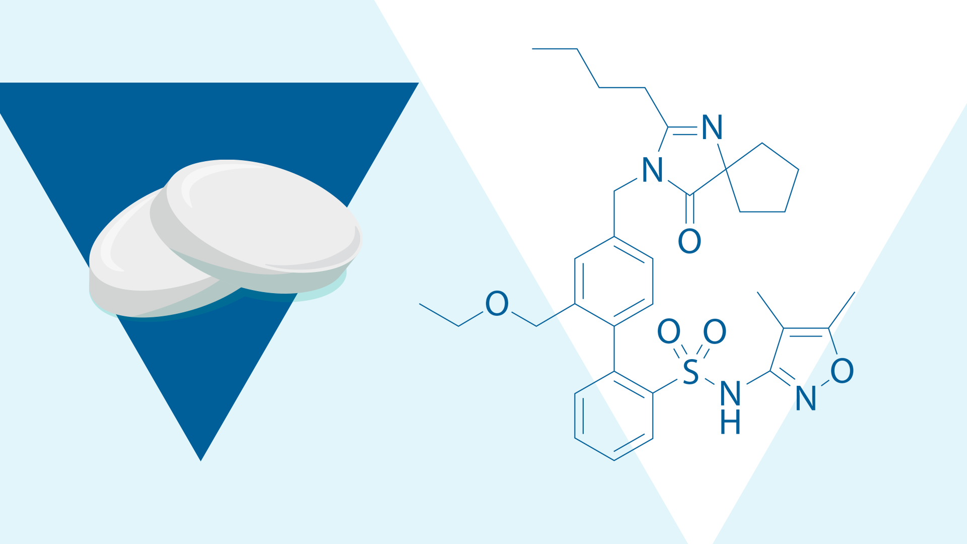
Sparsentan is a single-molecule, Dual Endothelin Angiotensin Receptor Antagonist (DEARA).8 Its mechanism of action is unique relative to other therapies used to treat FSGS.8
Using animal models that develop human-like FSGS can provide a greater understanding of the therapeutic mechanisms of sparsentan in FSGS.1
This study aimed to better understand the nephroprotective mechanisms of sparsentan compared to angiotensin receptor blockers (ARB) in a mouse model of FSGS using intravital multiphoton microscopy (MPM).1
Several transgenic mouse models were used, with equal numbers of males and females, including physiological (6-8 weeks old) and FSGS disease models (6 months to 1.5 years old).1
Three treatment groups of wild-type and transgenic mice were examined1:
Nephroprotective mechanisms were assessed by intravital MPM, enabling direct visualization of treatment effects on glomerular hemodynamics, podocyte and endothelial function, and tissue remodeling, comparing sparsentan with losartan in both healthy control and FSGS transgenic mouse models.1
ET-1 with or without Ang II was injected into the carotid artery of the pre-treated mice to assess glomerular hemodynamic changes caused by agonist-induced vasoconstriction.1
In both healthy and transgenic FSGS mice, sparsentan demonstrated a greater improvement in hemodynamics, podocyte and endothelial cell functions, and tissue repair compared to losartan.1
Hemodynamics and glomerular filtration
Compared to losartan, sparsentan1:
As a result, sparsentan more effectively reduced proteinuria and albumin leakage versus losartan.1
Podocyte and endothelial protection
Sparsentan attenuated ET-1 and Ang II-induced calcium spikes in podocytes and mesangial cells as well as restored endothelial glycocalyx and reduced immune cell homing.1
Tissue repair and regeneration
Both sparsentan and losartan decreased glomerulosclerosis and fibrosis, but sparsentan led to greater increase of podocyte number versus losartan.1
Additionally, sparsentan promoted renin- and endothelial-lineage clonal expansion in glomeruli and tubules and attenuated mitochondrial stress.1
This study used an animal model of FSGS to provide insight into the mechanistic actions and pleiotropic nephroprotective effects of sparsentan.1 Across multiple aspects of renal pathophysiology, the greater efficacy of sparsentan, a DEARA, compared with an ARB highlights the interplay of ET-1 and Ang II signaling in FSGS pathogenesis and management.1
Related Content
Mechanism of Protective Actions of Sparsentan in the Kidney: Lessons From Studies in Models of Chronic Kidney Disease
Clinical Science – 2024
This work was supported by a research collaboration grant from Travere Therapeutics Inc. Please see the publication for the full list of disclosures.
Ang II, angiotensin II; ARB, angiotensin receptor blocker; DEARA, Dual Endothelin Angiotensin Receptor Antagonist; ESKD, end-stage kidney disease; ET-1, endothelin-1; FSGS, focal segmental glomerulosclerosis; GFR, glomerular filtration rate; MPM, multiphoton microscopy; TRPC6, transient receptor potential cation channel, subfamily C, member 6.
MA-SP-25-0091 | October 2025